


Valgus deformation of the foot thumb is the most frequent acquired deformation of the foot, characterized by the diversion of the thumb out.
The pathology is bilateral in nature, mainly affects women over 35 years old.In most cases, deformation is accompanied by chronic inflammation of the joint bag and deformed arthrosis of pusnephalang joints, pronational deviations from the first metatarsal bone.
Causes of the disease.Why is it dangerous?
The etiological causes of Valgo's deviations from the foot in the foot have not yet been fully studied.There are several theories of occurrence:
- Vestigial theory.In the mid -nineteenth century, it was believed that only the models were subject to this deformation due to the use of model shoes at high and -tops, but during the study this pathology was found in men who wore flat shoes.
- The theory of primary muscle weakness - was refuted in a detailed study of the problem.
- The theory of weakness of the ligament apparatus and the lack of aponeurosis of the single - most scientists adhere to this theory.
There are several factors that lead to the debut of this pathology:
- Overweight increasing the load on the feet.
- Dystrophic changes related to age in the ligament joint apparatus.
- The weeks of heels are more than 5 cm, narrowed on the foot.
- Existing deformations of the skeleton (scoliosis, deformation of the femoral and knee joints valgus, flat feet).
The danger of pathology lies in the fact that, over time, this deformation leads to posture deformation, it is possible to develop dystrophic depths of the spine, the hip and knee joints due to the inadequate load distribution during walking and the injury of the foot muscles is accompanied by constants, which is significantly concerned the well.
Clinical manifestations
At the beginning of the disease, Valgus's deformation is not clinically.Most women are more concerned with the emergence of increased feet in the metacarpal-phalanx joint and the explosion of the first finger becomes noticeable.
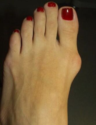
Over time, pain occurs, first when wearing narrow shoes and prolonged walking, and later the pain begins to be constant, painful.
Due to inadequate weight distribution, the member, for the first and second finger in the sole, is formed by Horn, the second finger is raised, strongly extended, the "Malleus" fingers are formed, which greatly complicates the walk.Due to the deterioration of blood circulation and innervation in the anterior part of the foot, arthrosis and chronic bursitis develop.
Diagnosis
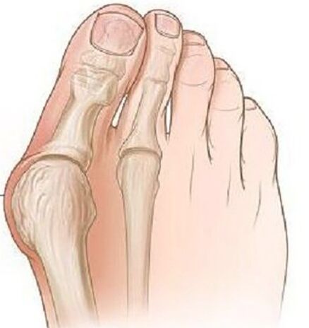
- During an objective exam, classic deformation with valgo deviations from the thumb is visible, the distal part of the foot has been expanded.In the projection of the head of the first metatarsal bone, there are signs of hyperemia and skin edema, pain during palpation.
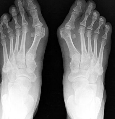
- The radiography of the foot in two projections.The frontal projection determines the degree of the thumb, the state of the metacarpal-phalanx joint, the degree of displacement of the sesamoid bone and, in the lateral projection, the degree of flat feet is visualized and calculated, which often occurs with the valgus deviations of the first toe.
- Plantation.In the print of the foot, made on paper, a line is designed by the center of the heel and between the fourth and third fingers, the external set of the foot is formed figuratively, according to which the presence of flattening of the foot and its degree is evaluated.In these patients, a flattened foot or straight feet of the 1st degree is most often detected.
Treatment and Prevention
Depending on the severity of the process, conservative or surgical treatment is performed.
Conservative treatment is performed at the mild stage of the disease, various types of orthopedic insoles are used, which are selected individually and bring the deformed finger to the normal position, while the load on the front of the limb is stabilized and distributed.
At the age of children and the senil, transverse bandage of the distal limb is used with a placement between the first and second finger.To reduce pain, hot baths with sea salt and soda are used, the radon baths give a good result.
To combat the bursitis, local anti -inflammatory gels are used, diexide and lidocaine compresses, and pronounced pain, novocaine block and intraarticular glycocorticoid injections.
Surgical treatment is possible at any stage of the disease, is performed under local anesthesia, which reduces the list of contrasts for surgery.
At the first stage, cold bone growths are removed on the inner edge of the bone, while it is only possible to reduce the pain of the process, but not to prevent the development of deformation in the future.
With a severe deviation of the finger with the presence of boring feet, an osteotomy is performed at the base of the positive bone of the first finger using a bone blade to reject the finger in the normal position, with the help of a Lavsan tape, the transverse sole ligament is formed.After surgery, the leg is strongly fixed with a dressing for 4 weeks, it is recommended to use individual orthopedic insoles during the year.
The prevention of the disease is to wear comfortable high heels, the shoes should sit on the leg freely, a tight toe should not cause discomfort in the thumb.If there is any deformation of the skeleton, it is advisable to undergo preventive examinations in the orthopedist to identify and slow the progression of the deformation of the foot in the foot.
Consequences and complications
A complication of the process is the development of Deichländer or "March" of the foot, which is characterized by acute pain, due to microcucks in the pairing joints of the foot and having vaginitis.
Due to constant inflammatory process and mechanical damage, there is a risk of developing malignant bone neoplasms.